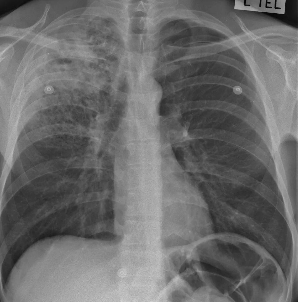
FIND: There is abnormal opacity in the upper half of the chest on the right. The rest of the image is normal.
Magnification of the right upper chest:

IDENTIFY: The upper part of the abnormal area is of the same density as the heart, except at its edge where it has a fluffy appearance (white oval). This means that at least part of the opacity is caused by airspace disease.
There is more fluffy opacity in the lower part of the abnormal area (yellow oval); this has small holes in it.
And within the dense upper area there is a cavity (blue oval).
LABEL: There is dense airspace opacity in the right upper lobe, containing a central cavity. Airspace opacity is also present in the right mid-zone.
MATCH: Given the imaging abnormality and the clinical history, the best match is active pulmonary tuberculosis.
SUMMARISE: Active tuberculosis in the right upper lobe.
Sputum assessment would be needed for absolute confirmation but the Xray appearances are virtually diagnostic. This is typical secondary, or re-activation, TB because of the upper lobe involvement and the cavity. The radiographer should confidently suggest the diagnosis.
Because the upper part of the lung is so densely opacified, we cannot be sure that there is no pleural disease next to it. But there is no fluid in the lower part of the chest, so there is at least no free pleural fluid. (if there was a large effusion causing shortness of breath, it might need to be aspirated; otherwise it would not change treatment).
The holes in the airspace opacity in the yellow oval are most likely due to underlying lung destruction caused by emphysema.