• Home
    • Abdomen
    • Chest 1
    • Chest 2
    • Head and neck
    • Skeleton
    • Abdomen
    • Chest
    • Head and Neck
    • Pathology introduction
    • Skeleton
  • Background
  • Contact
Menu

icxrays.org

  • Home
  • Cases
    • Abdomen
    • Chest 1
    • Chest 2
    • Head and neck
    • Skeleton
  • Topics
    • Abdomen
    • Chest
    • Head and Neck
    • Pathology introduction
    • Skeleton
  • Background
  • Contact

S1. Female, 25. Severe pain after minor trauma

April 6, 2017
In SKELETON Tags Bone tumour, Fracture, Pathological fracture

C3. Male, 34. Unwell with fever

April 5, 2017

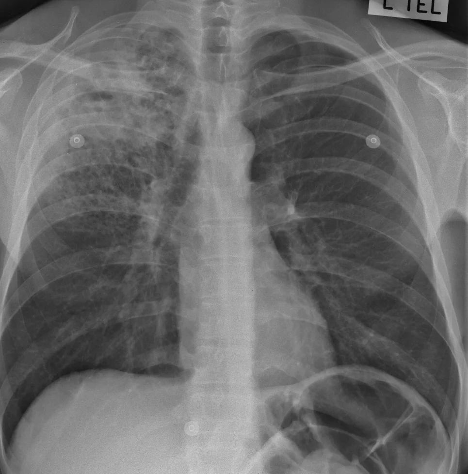
FIND: A huge number of tiny densities are scattered through both lungs. There is a circular dense line in the apex of the right lung. The right lung hilum is higher than normal. No other abnormality is seen. 

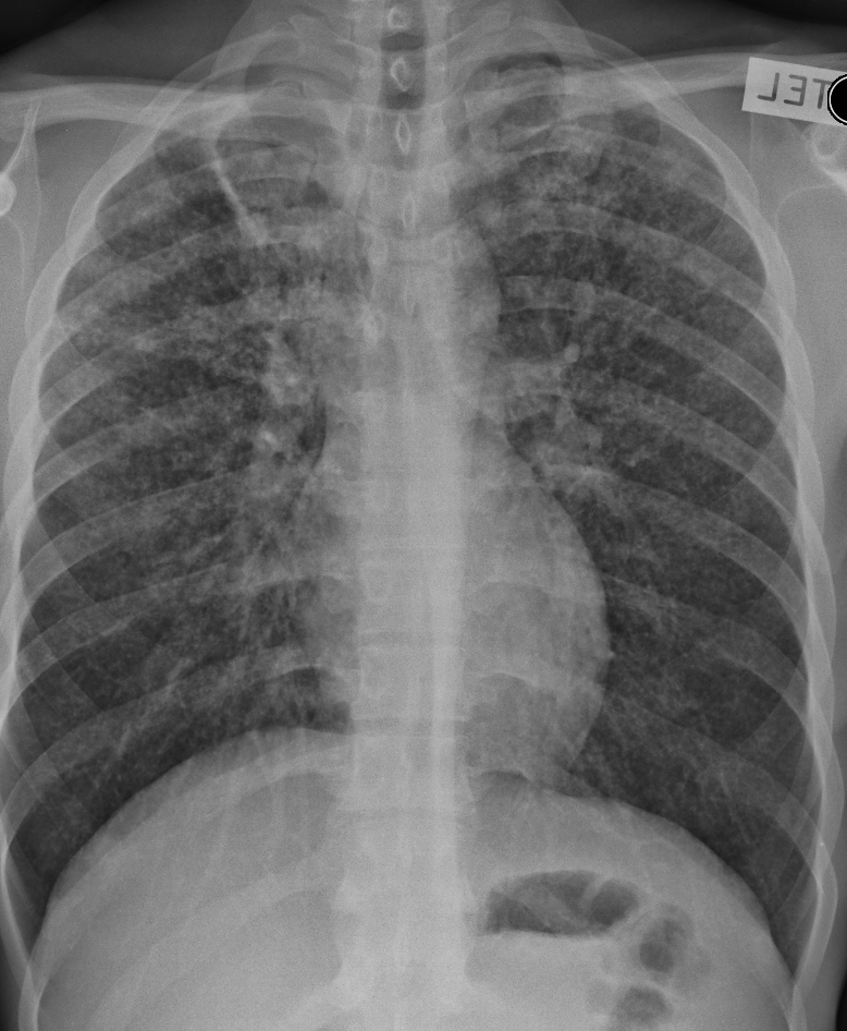
Here is a magnified view of the right upper lobe:

IDENTIFY: A close-up shows that the lung looks as if it is made of thousands of little dots, or nodules, one or two mm in size. There are so many of them that we can't see the vessels any more. And the abnormality near the right lung apex has  the appearance of a cavity with a smooth, thin wall. A high lung hilum usually means that it has been pulled up, either by a lobe which has lost volume or by scarring. 

LABEL: There are thousands of tiny nodules scattered through both lungs, and there is a cavity at the right lung apex. The right lung hilum is pulled up but there is no sign of collapse (or atelectasis) of the right upper lobe. So it's likely that there is some fibrous scarring, which we cannot see because of the nodules.

MATCH: In a patient with this history the lung nodules are almost certainly due to miliary tuberculosis. The cavity and the presumed lung scarring also fit with tuberculosis. If the patient was not very ill, the nodules could be due to cancer metastases (although this is very uncommon), with underlying changes of old tuberculosis.

SUMMARISE:  Miliary tuberculosis, with apical lung cavity likely due to tuberculosis as well. Right upper lobe scarring causing elevation of the right lung hilum.

Even though cancer is possible, TB would always have to be first on the list of diagnoses in an X-ray like this. Cancer would only be considered if TB could not be proven with sputum analysis and bood tests.

The right lung hilum is lower than the left hilum in 98% of normal people. 

Note that this case is a little unusual, in that military TB is usually seen in primary infection, but this patient is an adult; he also has a lung cavity which is a sign of secondary tuberculosis. This reminds us that diseases don't always follow the rules, especially TB which is changing in the era of increased drug resistance and the effects of HIV/AIDS. 

In CHEST Tags Cavity, Hilum, Miliary, Tuberculosis

C2. Female, 53. Cough and fever

April 4, 2017

FIND: There is an area of increased density in the lower part of the chest on the right. The rest of the image is normal. 

IDENTIFY: The abnormal area is uniformly dense, about the same as the heart, apart from a branching low-density area medially (yellow arrow) which follows the line of the right main bronchus (blue arrow). This is an air bronchogram.

The abnormal area  has a sharp upper margin which is almost horizontal (green arrow) .

The right border of the heart cannot be seen but the silhouette of the right side of the diaphragm (red arrows) remains visible. 

LABEL: The air bronchogram means that there is airspace disease present. The lost right heart silhouette indicates that the airspace disease is next to the heart, therefore it is anterior, therefore the middle lobe is abnormal; the sharp upper border is caused by airspace disease contained in the lobe by the horizontal fissure; and the visible diaphragm silhouette means that there is at least some normal lung against the diaphragm (therefore in the base of the lower lobe). 

MATCH: Airspace disease plus fever makes pneumonia the diagnosis, especially if the white cell count is raised. 

SUMMARISE: Right middle lobe pneumonia.

You don't need a lateral view to make the diagnosis but it is useful to understand the appearances in the frontal view. The middle lobe forms the dense white triangle superimposed over the heart, and you can imagine the beam running along its sharp upper border (near the hilum) to give the line shown by the green arrow. Also along the top of the curve of the diaphragm, just behind the base of the triangle anteriorly, to give that silhouette.

Why is there so much disease in just one lobe? It may mean there is something blocking the airway so that bacteria can't be expelled easily and have more chance to grow. There could be thick mucus in a child, or possibly cancer in an older person.

At any age, enlarged lymph nodes (TB, other infections or cancer) could squeeze the airway. Sometimes the airway (especially to the middle lobe) is narrowed by scarring. However sometimes it just happens like this!

Because there may be a treatable underlying cause, this patient should have a follow-up examination in 6 weeks' time, and unless the X-ray looks much better, further investigation might be needed. 

 
In CHEST Tags Fissures, Middle lobe, Pneumonia, Silhouette
C1.jpg

C1. Male, 25. Cough, haemoptysis and weight loss

April 3, 2017

FIND:  There is abnormal opacity in the upper half of the chest on the right. The rest of the image is normal.

Magnification of the right upper chest:

600.23 ivdu never confirmed. 1955036 0001.jpg

IDENTIFY: The upper part of the abnormal area is of the same density as the heart, except at its edge where it has a fluffy appearance (white oval). This means that at least part of the opacity is caused by airspace disease. 

There is more fluffy opacity in the lower part of the abnormal area (yellow oval); this has small holes in it.

And within the dense upper area there is a cavity (blue oval). 

LABEL: There is dense airspace opacity in the right upper lobe, containing a central cavity. Airspace opacity is also present in the right mid-zone.

MATCH: Given the imaging abnormality and the clinical history, the best match is active pulmonary tuberculosis.

SUMMARISE: Active tuberculosis in the right upper lobe. 

Sputum assessment would be needed for absolute confirmation but the Xray appearances are virtually diagnostic. This is typical secondary, or re-activation, TB because of the upper lobe involvement and the cavity. The radiographer should confidently suggest the diagnosis.

Because the upper part of the lung is so densely opacified, we cannot be sure that there is no pleural disease next to it. But there is no fluid in the lower part of the chest, so there is at least no free pleural fluid. (if there was a large effusion causing shortness of breath, it might need to be aspirated; otherwise it would not change treatment).

The holes in the airspace opacity in the yellow oval are most likely due to underlying lung destruction caused by emphysema. 

In CHEST Tags Airspace, Cavity, Tuberculosis